Earlier this year, Jamaica Hospital’s Radiology Department opened its new modernized mammography suite that utilizes state-of-the-art equipment offering advanced breast imaging services.
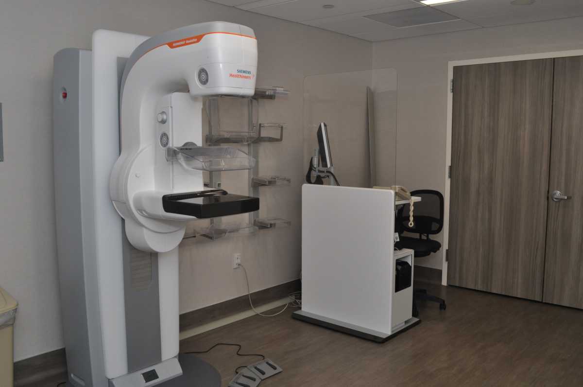
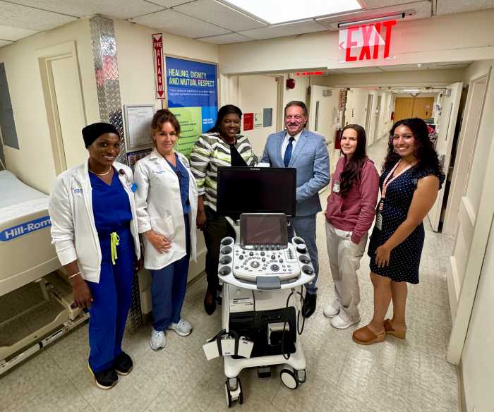
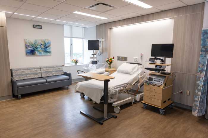
The new mammography suite, located on the first floor in the Department of Radiology, includes an ultrasound room, changing room, a handicap accessible bathroom and mammogram room where patients have been screened for breast cancer. The suite’s technology includes new Samsung ultrasound machines and a new Siemens 3D mammography machine (also known as a digital breast tomosynthesis).
A 3D mammogram is a low-dose diagnostic imaging procedure, which allows X-ray images of the breast to be captured from various angles and later combined to create a more comprehensive three-dimensional image of the breast.
In observance of Breast Cancer Awareness Month, Dr. Sohita Mehra, director of Breast Imaging at Jamaica Women’s Health Center and Flushing Hospital, wants women to know that they can come to the center that provides exceptional quality care to patients.
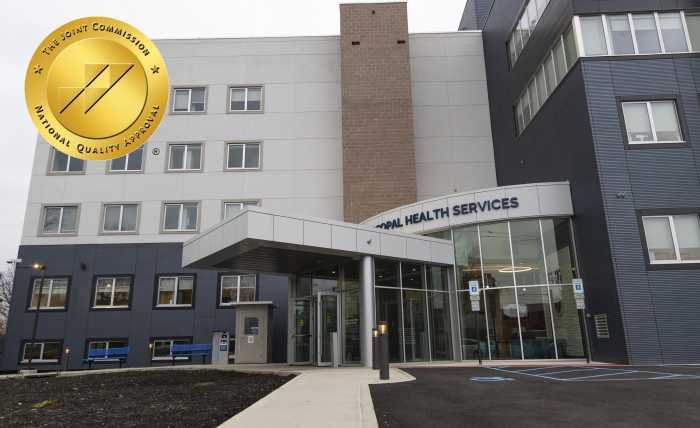
Jamaica Hospital, which is designated as a Diagnostic Imaging Center of Excellence by the American College of Radiology, upholds the highest standards of imaging quality with continuous standards in place to minimize radiation. All images are interpreted by board certified, expertly trained radiologists in breast imaging.
“Our technologists are so great. They’re used to seeing women of all shapes and sizes, different comfort levels and different personalities,” Mehra said. “They’re good at their jobs and they’re doing something meaningful. I, and the other doctors, are also here when they’re doing the procedure, and if they have any questions, we’re here to talk to them about it.”
According to Mehra, women who go in for an ultrasound are encouraged to do a mammogram as well, as it’s proven to detect breast cancer and help to prolong their lives.
“The reason why screening exams like this are important is we want to find breast cancer when it’s most treatable as possible and before you can feel one,” Mehra said. “If you’re over the age of 30 and feel a lump in your breast and have your doctor look over it, you can start with a mammogram and ultrasound. If you’re under 30 and, because your breasts are harder to see through, you’ll start with an ultrasound.”
The American College of Radiology recommends women should have the option to have a mammogram every year, starting at age 40 and every year after that.
“If you have first degree, relative with early breast cancer or you’ve received treatment like radiation for another type of breast-related cancer, then you would start earlier,” Mehra said. “You can talk to your doctor about early screening.”
While most people talk about the discomfort involved rather than the lifesaving possibilities associated with mammograms, Mehra said it’s actually not so unpleasant.
“Your breasts tend to be more tender before and during your period. If you can make an appointment right after, it’s less painful. The idea of the compression is that we can see better through your breasts,” Mehra said. “We get better imaging and we’re able to see more when doing the compression.”
The Radiology Department’s new 3D machine, according to Mehra, includes an auto sensor to not over-compress. The department has received good feedback from patients about their new updated suite.
“We do a really good job and if you need anything done further, we can get you to see the breast surgeons very quickly,” Mehra said. “We discuss cases on a weekly basis. You get great care at Jamaica Hospital.”
The hospital is working on producing a podcast that will discuss the mammography suite and will be shared on Facebook, Instagram and Twitter.

